

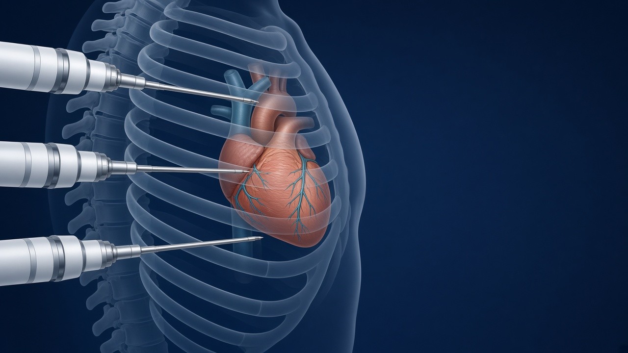
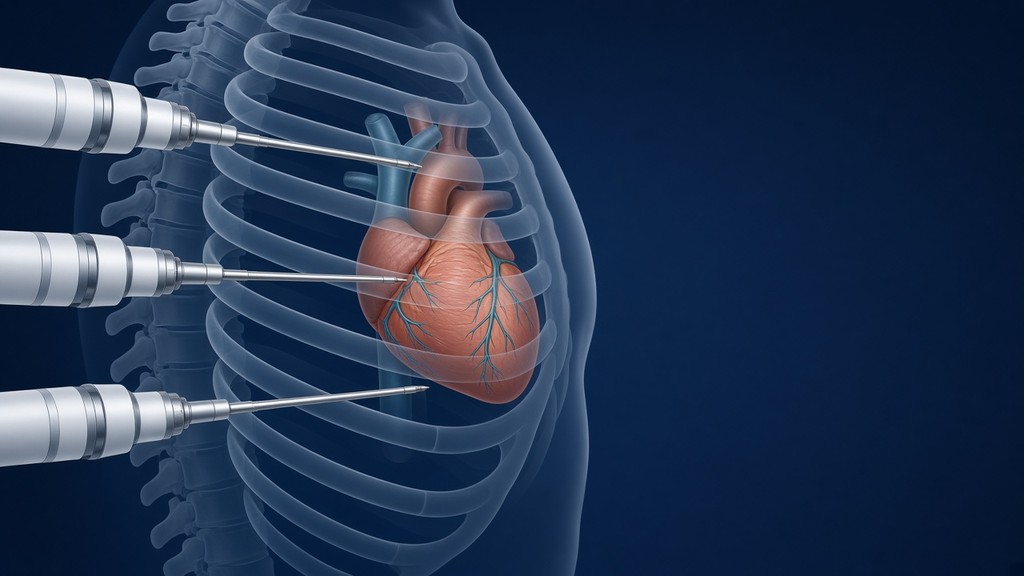
Robotic coronary surgery lets us perform a bypass without opening the breastbone, working through a few small openings between the ribs. We use the same system to repair the mitral valve and to close a hole between the upper chambers of the heart (an atrial septal defect, or ASD). In this article I want to walk you through what actually happens in the operating room, from preparing the bypass graft to the last stitch.
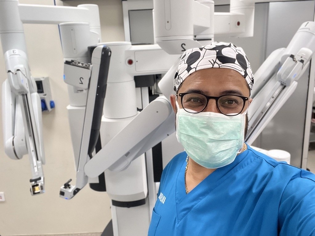
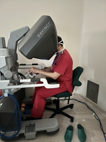
The question I hear most often in clinic is: “So, does the robot do the operation?” No. The robot does not make a single move on its own. I sit at the console and control the arms with my hands and feet, while the rest of the team stays at the patient’s side. The robot is a tool that magnifies what I see and lets me turn my wrist in spaces too tight for a human hand.
Please note: This article is for general information only and does not replace an examination, diagnosis or treatment decision. If you have sudden chest pain, shortness of breath or feel faint, seek emergency care right away.
I have covered why and for whom robotic surgery is chosen in why use a robot in heart surgery, and what the system adds to bypass surgery in what does da Vinci add in robotic bypass surgery. Here we go one step further, into the operating room itself.
Who stands where in the operating room?
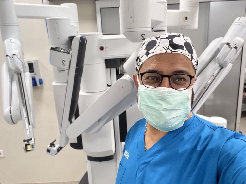
Robotic surgery is a team effort. The robot’s arms are positioned at the operating table, and the camera and instruments attached to them enter the chest through ports about 8 millimetres wide. At the table are our assistant surgeon, scrub nurse, anaesthesia team and, when needed, the perfusionist who runs the heart-lung machine.
I am in the same room, a few metres away at the console. Looking into it, I see the heart in three dimensions, magnified roughly ten times. The movements of my fingers are transferred to the arms. The system filters out hand tremor and can scale a large hand movement down to a millimetre-level instrument movement. Because the instrument tips bend and rotate like a wrist, we can place stitches comfortably in corners that are hard to reach even with an open chest.
At every stage we are set up to convert to conventional open surgery if needed. It is rarely necessary, but it is part of the plan. Safe robotic surgery depends on a team that can make that switch without hesitation.
Step one in robotic coronary surgery: preparing the graft
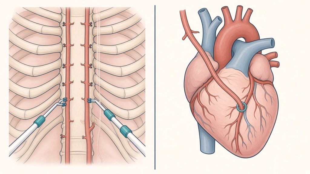
In a bypass operation we connect a new blood supply beyond the blocked coronary artery. The most valuable vessel for this is the internal mammary artery (also called the internal thoracic artery, or LIMA), which runs along the inside of the chest wall just beside the breastbone. When it is connected to the left anterior descending artery (LAD), it stays open for many years in the vast majority of patients, which is why it is the graft surgeons trust most. You can read more about graft options in where do bypass grafts come from.
In open surgery, reaching this artery means dividing the breastbone and lifting the chest wall with a retractor. With the robot, we proceed like this:
- Together with the anaesthesia team, we temporarily deflate the left lung to create working space inside the chest.
- We place the camera and two instruments through three small ports on the left side of the chest.
- We free the artery from the inner surface of the chest wall, working from the level of the collarbone downwards and clipping its side branches one by one. The magnified view makes it easier to handle this delicate vessel without injuring it.
- We check the length and flow of the artery and mark the spot on the heart where it will be connected.
How is the bypass connection made?
Once the graft is ready, there are two ways to make the connection (the anastomosis). Which one we choose depends on the patient’s anatomy and the condition of the heart.
- Through a small incision (robot-assisted MIDCAB): We make an incision of about 4–5 cm between the ribs, below the left breast. With the heart still beating, we steady the target area with a special stabiliser and sew the connection directly. Most patients do not need the heart-lung machine.
- Totally endoscopic bypass (TECAB): The connection itself is also made with robotic instruments, through the ports. This is used in selected patients.
Either way, we measure flow through the graft after the connection is made. We want to see that the bypass works well before the patient leaves the operating room.
If more than one artery is narrowed, hybrid treatment may be an option: the LIMA is connected to the LAD robotically, and the cardiology team places stents in the other arteries. This is a decision made jointly by the cardiologist and the heart surgeon. I discussed the differences between the two in stent or bypass. For many patients with severe blockages in all three main arteries, conventional open bypass is still the best choice.
Robotic mitral valve repair
Mitral valve repair is the area with the longest track record in robotic heart surgery. The mitral valve is the door between the left atrium and the left ventricle. When one of its leaflets prolapses, or the cords that hold it in place (the chordae) rupture, the valve no longer closes properly and blood leaks backwards. Over time this can enlarge the heart, trigger rhythm problems and cause breathlessness.
Valve surgery means working inside the heart, so we connect the patient to the heart-lung machine through a small incision in the groin. We enter through a few small ports and a working incision of about 3–4 cm on the right side of the chest, stop the heart and open the left atrium. We then see the valve face-on, magnified and in three dimensions. The repair usually involves these steps:
- Identifying the part of the leaflet that is prolapsing or has a torn cord.
- Where needed, removing excess tissue or placing thin artificial cords (ePTFE sutures) that anchor the leaflet to the heart muscle.
- Bringing the stretched valve opening back to its normal size with a ring sewn around it (an annuloplasty ring).
- Testing the valve by filling the ventricle with saline, then confirming the result with transoesophageal echocardiography in the operating room once the heart is beating again.
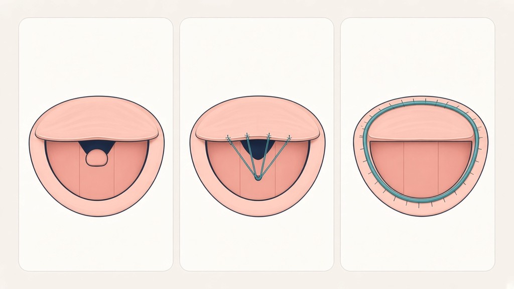
Repairing a valve is usually better than replacing it. The patient keeps their own tissue, and the lifelong blood thinners required with a mechanical valve are usually not needed. When repair is not possible, the valve can be replaced through the same approach. Tricuspid valve repair, closure of the left atrial appendage or ablation for atrial fibrillation can also be added during the same operation.
ASD closure: a hole between the upper chambers
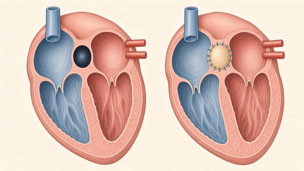
An atrial septal defect (ASD) is a hole in the wall that separates the right and left atria. It is present from birth and is often found in adulthood, when palpitations, tiredness or an echocardiogram done for another reason bring it to light. The extra blood crossing the hole gradually strains the right side of the heart and the blood vessels of the lungs.
Many ASDs can be closed without surgery, using a closure device placed through a vein in the groin. Surgery is needed when the hole is very large, when its edges are not strong enough to hold a device, or when it sits in a location unsuitable for a device, such as a sinus venosus defect. This is one of the situations where the robotic approach makes the most sense: these patients are often young and active, and we can carry out the same repair without opening the breastbone.
The operation is done from the right side of the chest, much like mitral valve surgery. With the patient on the heart-lung machine, we open the right atrium and see the hole directly. Small holes can be closed with stitches; larger ones are closed with a patch we prepare from the patient’s own pericardium (the sac around the heart). If a sinus venosus defect comes with lung veins draining to the wrong chamber, we redirect that flow to the correct atrium in the same operation.
Who is not a good candidate for robotic surgery?
The robotic approach is a powerful tool, but it is not the best option for every patient. We usually choose a different route when there is:
- Previous chest surgery on the same side, or extensive scarring of the lining of the lung after severe pneumonia, tuberculosis or similar conditions
- Lung disease so advanced that the patient cannot tolerate breathing with one lung during surgery
- Severe calcification of the groin and leg arteries or of the aorta, because the heart-lung machine is connected through these vessels
- Severe disease in several coronary arteries, where complete open bypass is the safer choice
- An emergency, an ongoing heart attack or very poor heart function
We make this decision based on the examination, echocardiography, coronary angiography and, when needed, a CT scan. Sometimes the most honest thing I can tell a patient is, “For you, conventional surgery is the safer option,” and I do not hesitate to say it.
What to expect after surgery
Because the breastbone is not divided, there is no bone to heal, which is often the hardest part of recovery after open surgery. Most of our patients come off the breathing machine shortly after surgery, are walking the next day and usually go home within a few days. Returning to desk work and driving is typically weeks earlier than after open surgery; we discuss the timing with each patient individually. The incision between the ribs can be sore and tender for the first few weeks, and this is managed with pain medication.
The operation may be different, but the care afterwards stays the same: taking medication regularly, controlling cholesterol and blood pressure, quitting smoking and joining a cardiac rehabilitation programme. I cover the weeks after bypass in detail in recovery after bypass surgery.
Frequently asked questions
Does the robot perform the surgery?
No. Every movement of the robot is controlled by the surgeon at the console. The system does not make decisions, place stitches or cut on its own.
Does robotic surgery take longer?
Setting up and positioning the arms can make the operation somewhat longer than open surgery. In return, the hospital stay and the time it takes to get back to daily life are often shorter.
What kind of scar will I have?
There is no long incision down the middle of the chest. You will have an incision a few centimetres long on the side of the chest and a few small port scars. In women, we can often hide the incision in the crease beneath the breast.
Can the operation be converted to open surgery?
Yes. It is rarely needed, but our team is prepared for it in every robotic operation. The patient’s safety comes before the size of the incision.
Are the results of robotic bypass the same as open bypass?
In the right patient and in experienced hands, the long-term results of a LIMA-to-LAD bypass are comparable to those of open surgery. What matters most is not the robot, but choosing the right patient and the quality of the connection.
If you would like to find out whether robotic coronary surgery, mitral valve repair or ASD closure is suitable for you, you can book a consultation and bring your test results with you.