


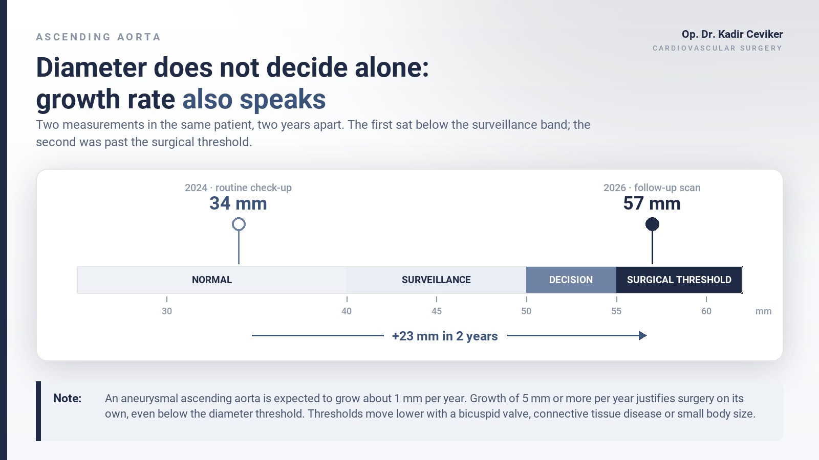
I am writing this with my mother’s permission. In 2024, during a routine cardiac check-up, one line at the bottom of her echo report read: ascending aorta 34 mm. Nobody raised an alarm, and honestly nobody had to — that number sits right at the border. In 2026 I scanned her myself. The same vessel measured 57 mm.
She had no complaints at all. The only new finding was blood pressure that had recently climbed well above target.
A note before we start: this is general medical information, not a personal treatment plan. If chest pain starts abruptly, tears through to the back and peaks within seconds, call emergency services immediately.
What is an ascending aortic aneurysm?
When the first segment of the main artery leaving the heart widens well beyond its normal calibre, we call it an ascending aortic aneurysm — the “ballooning artery” patients have heard about, only in the chest, a few centimetres from the heart.
In an adult woman the ascending aorta usually measures around 30 mm. Past 45 mm we are talking about an aneurysm; 50–55 mm is the band where surgery enters the conversation. I covered aneurysms in general in the aneurysm article; here the rules differ from the abdominal aorta.
When an abdominal aorta ruptures, the bleeding is often contained long enough for some patients to reach an operating room. In the ascending aorta the bigger threat arrives before rupture: aortic dissection. The layers of the wall separate and blood tracks into the wrong plane. Untreated type A dissection is an emergency where risk climbs by the hour over the first two days.
23 millimetres in two years — is that normal?
No. It is nowhere near normal, and it is the reason I am writing this.
An aneurysmal ascending aorta usually behaves slowly: roughly 1 millimetre of growth per year. That is why we plan follow-up in years rather than months. My mother’s aorta moved about eleven times faster — close to 11 millimetres a year. The line at which guidelines say a vessel can no longer wait is 5 millimetres per year, so she was double that on growth alone, on top of an already surgical diameter.
I hear this in clinic constantly: “The echo said mild dilatation, but my doctor didn’t mention it.” Most of the time there genuinely is nothing to do that day. But “nothing to do today” and “never look again” are not the same sentence. Every borderline aorta deserves a date for the next scan before the patient walks out.
Why did it cause no symptoms?
Because a widening aorta does not hurt. There are no pain fibres to trigger, and until it presses on a neighbour it stays silent.
A very large ascending aorta can stretch the nerve to the vocal cord and cause hoarseness, push on the airway and produce a stubborn cough, or tug on the aortic valve and make it leak — which shows up as breathlessness and fatigue.
But in most patients there is no warning at all. The first symptom is the dissection itself: a tearing pain in the middle of the chest that peaks within seconds and radiates to the back. Unlike a heart attack it does not build gradually — it arrives at full volume.
When I took my mother to theatre she still had no symptoms. That is exactly why we could operate electively, on a planned morning list. This is not a footnote to the story; it is the whole story.
What I saw in the operating room: diameter is not the full picture
We ( with Prof. Deniz Şerefli) performed an ascending aortic graft interposition — the dilated segment was excised and replaced with a synthetic graft. The operation was completed successfully.
But the vessel in my hands looked worse than the vessel on the scan. The middle layer of the wall, the media, was severely thinned and carried widespread atherosclerotic change; in places it was close to paper-thin. Let me be blunt: that aorta was very near a complication. One hypertensive surge, one heavy strain, a few more weeks of waiting might have been enough.
This is what imaging cannot tell us. A CT scan gives diameter to the millimetre. It does not report how much strength is left in the wall.
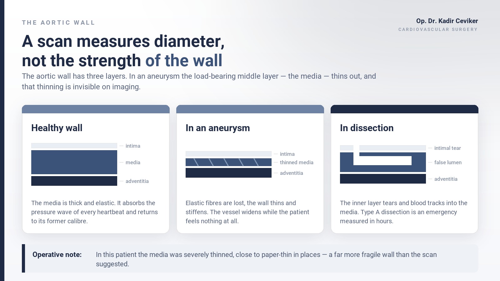
International dissection registries make the same point from the other direction: more than half of patients who suffer a type A dissection had an aortic diameter below the classic 55 mm threshold. The threshold is a decision aid, not a guarantee of safety.
Where blood pressure fits in
My mother developed significant hypertension over the same period. That is not a coincidence. The aorta absorbs the pressure wave of every single heartbeat; when blood pressure rises, wall stress rises with it, and a media that is already thinned fatigues faster. Uncontrolled hypertension both accelerates aneurysm growth and raises dissection risk.
So in a patient with an aneurysm, antihypertensive treatment is not a “let’s watch it for now” topic. Starting medication does not make the artery dependent on the drug — I addressed that common fear in the piece on whether blood pressure medications cause addiction. Smoking belongs on the same list; it directly undermines the wall’s capacity to repair itself.
When do we operate, and when do we watch?
Three things drive the decision.
Diameter. For a degenerative aneurysm, 55 mm is the classic threshold. With a bicuspid aortic valve, a connective tissue disorder or a strong family history, it drops to 50 mm and lower in selected patients. In small or slender patients we index diameter to height or body surface area rather than trusting the absolute number, and for the same diameter the risk runs higher in women.
Growth rate. An increase of 5 mm or more per year justifies surgery even if the diameter has not reached the threshold. That was my mother’s situation — she met both criteria.
Associated problems. Significant aortic valve disease, another planned cardiac operation, or symptoms from compression all move the decision earlier.
One more factor: who operates. These thresholds are written on the outcomes of high-volume aortic centres.
Elective and emergency are not the same operation
If you take one sentence from this article, take this one: the same procedure is a completely different proposition planned than it is after the aorta tears.
Elective ascending aortic surgery in an experienced centre is a low-risk open heart operation. The patient walks in on the morning of surgery, spends a short stay in intensive care and on the ward, then goes home. When the same vessel dissects, the patient arrives at 3 a.m. — different mortality, different recovery.
My mother was in the first group. The only reason was that a 34 mm note from two years earlier had not been forgotten.
What if aortic aneurysm runs in your family?
Ascending aortic aneurysms cluster in families. Once one person is diagnosed, I recommend imaging at least once for first-degree relatives — parents, siblings, children. Echocardiography comes first; when it cannot show the whole ascending aorta, CT or MR angiography is needed.
If there is a bicuspid aortic valve — two leaflets instead of three — this screening is not optional. Dissection at a young age, several affected relatives or Marfan-type features bring genetic evaluation into play, which I covered in the article on heritable aortopathy and genetic testing.
If you are holding a borderline aortic measurement and nobody has told you when to repeat it, you can request an appointment from the contact page.
Frequently asked questions
My aorta measured 40 mm. Will I need surgery?
Most likely not. 40 mm sits in the surveillance band, not the surgical one. What matters is that the date of the next scan is set and that your blood pressure stays at target.
Can medication shrink an aneurysm?
No. The goal of medical treatment is to slow growth and reduce dissection risk. Tissue that has already stretched does not return to its old shape.
Can I exercise?
Walking, swimming and cycling at a steady pace are generally fine. Heavy lifting, maximal straining and anything done while holding your breath are not advised, because they spike blood pressure within seconds.
Is life normal after surgery?
Yes. The graft is permanent and does not wear out, and most patients are back to daily life within a few weeks. Blood pressure control and annual imaging of the rest of the aorta continue.
Is every chest pain a dissection?
No, and the vast majority are not. But pain that peaks within seconds, feels like tearing and radiates to the back is a different animal. With that description there is no waiting — call emergency services.
Scientific sources
- Isselbacher EM et al. 2022 ACC/AHA guideline for aortic disease (surgical thresholds, family screening) — Circulation, 2022
- Mazzolai L et al. 2024 ESC guidelines for peripheral arterial and aortic diseases — European Heart Journal, 2024
- Pape LA et al. IRAD: most type A dissections occur below 55 mm — Circulation, 2007
- Davies RR et al. Yearly rupture or dissection rates for thoracic aortic aneurysms by size — The Annals of Thoracic Surgery, 2002
WordPress publishing notes
- Category: blog (#238) / TR: Bloglar (#28)
- Featured image:
01-featured-aort-cap-skalasi-en.jpg· in-body figure:02-aort-duvari-media-diseksiyon-en.jpg - Rank Math focus:
ascending aortic aneurysm - Internal links: aneurysm-symptoms-follow-up-treatment, do-blood-pressure-medications-cause-addiction, heritable-aortopathy-genetic-testing-aortic-aneurysm, contact